rTMS Motor Threshold and Dosing Basics for Clinicians
rTMS dosing is individualised. Each patient’s cortical excitability differs, so treatment intensity is set relative to their motor threshold.
What is motor threshold?
The resting motor threshold (MT) is the minimum stimulator output that produces a motor evoked potential (or visible twitch) in a relaxed target hand muscle in about half of the attempts, commonly 5 out of 10.
Finding the motor hotspot
- Place the coil over the left motor cortex with the handle pointing backwards and about 45° from the midline.
- Stimulate at a moderate intensity while moving the coil in small steps.
- The hotspot is where the strongest, most consistent response appears in the target muscle (APB or FDI).
Determining MT
- EMG method. Record MEPs with surface electrodes; MT is the intensity that gives MEPs of at least 50 µV in about half of trials.
- Visual method. Watch for a thumb or finger twitch. It is practical but slightly less precise.
- Start above threshold and lower in small steps, then confirm.
Setting treatment intensity
- 10 Hz left DLPFC (depression): commonly 120% MT.
- Theta Burst: protocol-specific, often expressed relative to MT.
- Low frequency: protocol-specific.
Always follow the protocol your clinic has adopted and published safety limits.
Locating the DLPFC
Methods include the traditional "5 cm rule" (now considered less accurate), scalp-measurement methods based on the 10-20 system (e.g., the Beam F3 method) and neuronavigation. Use the same method for every session.
Consistency tips
- Mark coil positions or record measurements.
- Re-check MT periodically and after medication changes.
- Keep the patient relaxed during MT measurement.
- Record everything in the session log.
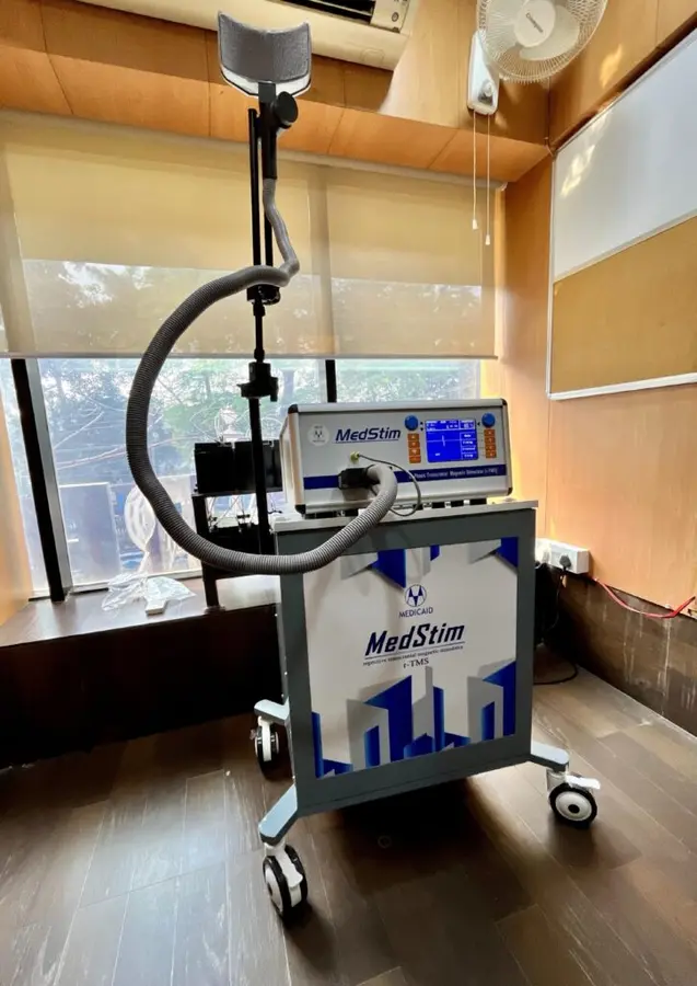
The protocol libraries on the MS-50 and MS-100 store parameters for consistent delivery. RS Enterprises includes MT and targeting training at installation.
MS-50 rTMS Machine
Indigenous rTMS with Theta Burst, 0.5–50 pps and dual coil support. ₹19 lakh – ₹24 lakh
Frequently Asked Questions
Which muscle is used for MT?
Commonly the abductor pollicis brevis or first dorsal interosseous of the hand.
Visual or EMG method?
EMG-based MT is more precise; visual observation of thumb twitch is widely used in clinics.
How often should MT be re-measured?
Many clinics re-check weekly or after medication changes.
This article is for general information for healthcare buyers and professionals and is not medical advice. Clinical decisions should be made by qualified practitioners.